The physiology of the bends

Gases and physiology go hand in hand. We breathe oxygen and exhale carbon dioxide. Several molecules that we usually come across in gas form act as signal molecules, for example carbon monoxide and nitric oxide. But sometimes, things go wrong when gases meet physiology. In this blog post, I will talk about one such situation: decompression sickness, also called the bends.

diving_free_stock

What, exactly, is the bends? The bends happens when a person has been under pressure and then that pressure is removed quickly, causing the gases that are normally dissolved in the body fluids to form bubbles. A typical situation is where someone has been diving deep underwater and then resurface too quickly*.

What causes it? The first thing we need to know is which gases are problematic, namely inert gases. There are inert gases in the body. An inert gas is a gas that, under the given conditions, does nothing – i.e. it doesn’t undergo chemical reactions. The inert gases in the body mostly sits there and does not take part in metabolism. Oxygen is not an inert gas in the body – it contributes to several physiological processes including respiration. Nitrogen, however, is an inert gas in the body – it does not contribute. It is the main inert gas in the air, and the most common culprit of the bends.

Nitrogen and other inert gases are typically dissolved in the bodily fluids. This process requires pressure. Henry’s Law describes the relationship between pressure and a gas dissolved in liquid. This law states that when pressure on a liquid is decreased, the amount of gas dissolved in the liquid will decrease to the same extent, and when pressure is increased, the amount of gas dissolved will increase. This means that if the human body is put under high pressure (e.g. when diving), the inert gases will be dissolved in the bodily fluids. This is not a problem. The gases are inert, doing nothing, so we can usually handle the extra amount. The problem occurs when the pressure is reduced, and it is reduced fast. If a diver is moving slowly from the high-pressure underwater environment to the surface, the gases being released may escape bit by bit as the person breathes out, which is fine. But if it happens fast, the lungs may not be able to keep up, and that causes trouble. The inert gases become un-dissolved in places where they really should not be: we get bubbles trapped in the body.

Bubbles are bad news. They disrupt blood flow and can cause damage. Bubbles can form in (or be transported to) any part of the body (although some places such as large joints are more at risk). This means that we can get a great range of symptoms, depending on where the bubbles form. The worst outcome is death, which can happen if the bubbles for example interrupt spinal cord function. Less severe outcomes, although still severely unpleasant, are seizures, paralysis, dizziness, pain, visual disturbances, breathlessness, nausea, incontinence and more. In short, we do not want bubbles of inert gas (usually nitrogen) interfering with our organ function.

The deeper and longer the dive, the more dangerous it can be. We know that pressure increases as you descend. At 10 meters, the pressure has increased from 1 atm (sea level) to 2 atm, and the same level of pressure will therefore be needed for the diving suit to remain inflated. For reference, one atmosphere is the pressure that the air exerts at sea level, and the pressure tends to increase with 1 atm for each 10 m increase in depth. In other words, each 10 m below the surface adds to the body of water pressing down on the diver. The further down, the more weight, and this weight is in addition to the weight of the air (1 atm at sea level). 

At 1 atm (sea level), the oxygen content is approximately 21%, and nitrogen content is approximately 78%. The oxygen pressure is therefore 0.21 * 1 atm =0.21 atm, and the nitrogen pressure is 0.78 * 1 atm = 0.78 atm. There are other pressure units that this can be measured in, such as mmHg or Torr (1 atm = 760 mmHg = 760 Torr) and kPa (1 atm = 101.325 kPa), but we will stick to atm for now.

If a diver is given normal air (with the percentage composition described above), at 10 meters the oxygen pressure will be 0.21 * 2 atm = 0.42 atm, and nitrogen pressure will be 0.78 * 2 atm = 1.56 atm. At this pressure, the blood will start to dissolve the nitrogen. This means that the fluids in the body will contain increasing amounts of nitrogen until an equilibrium is reached. How much nitrogen ends up where in the body depends on the tissue composition. Fat, for example, can dissolve a lot of nitrogen, and will take a bulk share.

Dissolving nitrogen is a slow process, as is releasing dissolved nitrogen. The longer the dive, the more nitrogen is dissolved, and the longer it takes to release it afterwards.

So why nitrogen? Why not oxygen, or carbon dioxide? As a rule of thumb, for the bends to happen, the diver has to experience the gas in question at a pressure of at least 2 atm. This excludes both carbon dioxide and oxygen. Even at depth, carbon dioxide pressure simply does not increase much above its normal level (about 40 mmHg, or around 0.05 atm), and oxygen is used up by the tissues so it does not rise as much either*. Unless the diver uses a gas mixture with a different inert gas (e.g. helium), nitrogen will be the villain of the piece.

There are advantages to using other gases. Helium, for example, is less soluble than nitrogen. This means it dissolves faster into the body fluids, but it is also removed faster, reducing the risk of developing the bends. For long dives, this is good, but for short ones (where nitrogen simply would not have time to dissolve much), it’s not so good. One may ask why use an inert gas at all, if they are so problematic, and the simple answer is that pure oxygen under pressure is toxic**.

What about diving animals? Plenty of animals have an aquatic mode of life, and the list includes a variety of species from the odd rodent, ungulate and marsupial to penguins, seals and whales. Many of these dive to incredible depth, and they ascend and descend quickly.

diving depths
Diving depths of marine mammals, not including the deepest diving whale (Cuvier’s Beaked Whale, 2992 m) or birds (deepest diving is Emperor Penguin at 565 m, and deepest diving flying bird is Brünnich’s guillemot at 210 m). The personal record of this particular mammal is 3 m, which was a proud, yet unpleasant, moment. *Record by Soviet submarine K-278 in 1984, data for current submarines classified. Image not to scale: the submarine was 117 meters long, making it around 4 times the size of a blue whale.

At depth, the lungs of diving animals are almost entirely collapsed and blood flow to the lungs limited. Some diving animals even have collapsible rib cages that forces the air out when they dive. This means that no nitrogen can enter the blood during the dive. Many diving animals have quite small lungs, possibly because of this (and because large lungs work as airbags, making diving difficult). The diving animals do not store oxygen in their lungs for their dives, but rather in their blood, and they are very particular in how they use what little oxygen they have.

Oxygen storage. Diving animals usually have much more blood relative to their body size than non-diving animals, and the blood itself has a greater oxygen capacity (almost 40 ml oxygen per 100 ml blood, twice that of humans). The increased oxygen carrying capacity is due to a greater amount of red blood cells (which contain haemoglobin, the molecule that binds oxygen), and a much higher level of haemoglobin in the muscles (called myoglobin). The sperm whale, for example, has a myoglobin content almost 10 times that of humans, allowing them to store a vast amount of oxygen. Theoretically, the high level should cause the myoglobin to clump together, causing disease, but diving animals’ myoglobin is positively charged and stays nicely separated even when the muscles are packed with it.

Oxygen use. This impressive store of oxygen, however, is not enough. In addition, most diving animals have a lowered heart rate during dives, and blood flow to most of their internal organs is lowered (the exception being the brain). Even the heart gets less blood (but then again, the heart rate is lowered, so less blood is needed). Muscles also get reduced blood flow despite being needed to swim – instead, they rely on anaerobic (non-oxygen) metabolism after the myoglobin oxygen storage is depleted. Many diving animals also reduce muscle use. For example, Weddell and Elephant seals, bottlenose dolphins and blue whales have all been shown not to use their muscles when descending: sinking rather than swimming. And diving animals also reduce their metabolic rate, meaning that they use less energy. This is made easier by many diving animals being large, because oxygen consumption relative to body size is smaller the larger the body size. In other words: a mouse uses much more oxygen per gram of its body (1.65 litre oxygen per kilo per hour) than does an elephant (0.07 litre oxygen per kilo per hour).

In short: diving animals avoid the bends through minimizing the amount of nitrogen entering their blood when they dive***. To stay submerged, they are very good at storing oxygen in their blood (through higher blood volume and greater amounts of red blood cells) and muscles (through greater myoglobin levels), and use less oxygen (through shutting off blood flow to non-important organ systems, reducing their heart rate and metabolic rate, letting their muscles work anaerobically or not work at all).

*Interesting fact: The bends can also happen when ascending rapidly in for example an aircraft, with the danger arising as the pressure is quickly reduced to 0.5 atm. The underlying principles remain the same. A pressurised cabin will prevent this entirely (as with deep-diving submarines).

**Oxygen at pressures above 1 atm is toxic. When diving deeper than 60 meters, the pressure is so high that the gas mix needs to contain less oxygen than at sea level (hypoxic mixture) to avoid oxygen poisoning. There are multiple studies on oxygen toxicity, but the wikipedia article on the topic is quite extensive and accessible: https://en.wikipedia.org/wiki/Oxygen_toxicity

***This does not mean that they cannot get the bends. For example, stranded whales have shown signs of gas-bubble tissue damage (Gas-bubble lesions in stranded cetaceans: Nature 425, 575–576 (2003) https://www.nature.com/articles/425575a). It is possible that this has to do with the animals being stressed (and therefore either diving for too long or ascending or descending too quickly), or having too high blood flow (again due to stress or possibly also cold). While we do not know the exact reason why and how this happens, we do know that the use of sonar has been linked to whales getting the bends.

Breathing on the brain

As easy as breathing? It may seem like it, but breathing is actually no easy process. It involves the precise integration of several systems, including, of course, the pulmonary and cardiovascular systems (gas exchange and transport, respectively). However, it also requires the direct involvement of the central nervous system: the brain.

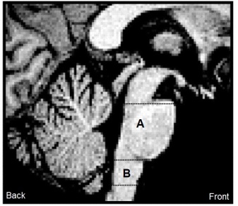
The drive to breathe is generated in the brainstem. In a study published almost 100 years ago, the crucial role of the different parts of the brainstem was demonstrated in a somewhat gruesome way by sectioning the brainstem of an anaesthetised cat (Lumsden, 1923). The main areas of interest are in the medulla and pons. Cut above this, and the breathing continue. Cut below this, and it will cease.

brainstem_small2

The brainstem is the part of the brain that connects to the spinal cord. The figure above shows an MRI image of a human brainstem, with the pons (A) and medulla (B) labelled.

The Medulla

On each side of the medulla we find neurons that are grossly organised in vertical columns. These can be divided into dorsal and ventral columns – that means columns situated towards the back of the medulla, and columns that are towards the front.

On the dorsal side of the medulla (towards the back), we find neurons that primarily govern inspiration (as opposed to expiration). These neurons fire when you breathe in. One of the most important clusters of neurons in this area is the nucleus tractus solitarius (NTS), which is an area that receives sensory input from the chest. It is thought that the NTS integrates sensory information from the body and relays this to other regions in the brainstem. In short, it could be viewed as a communication hub. The NTS is involved in a range of important functions as well as respiratory control, including for example the control of blood pressure.

On the ventral side of the medulla (towards the front), we find both inspiratory and expiratory neurons. We also find motor neurons that connect with muscles in the throat and chest. The respiratory column on the ventral side can be grossly divided into three regions: rostral (top/front), intermediate (middle) and caudal (bottom//back). The rostral part holds expiratory neurons. The intermediate part contains mostly inspiratory neurons plus the assumed ‘pacemaker’ of breathing: the pre-Bötzinger complex*. This complex contains pacemaker neurons capable of setting a breathing rhythm, and it can generate different rhythms depending on the level of oxygenation. We don’t yet know if the pre-Bötzinger complex sets the pace on its own, or if uses input from other regions as well. Typically, if not influenced by other factors, this region is ‘on’ for two seconds (inspiration) and ‘off’ for three seconds (expiration). Finally, the caudal regions hold expiratory neurons (that connect with motor neurons controlling expiration).

372px-2327_Respiratory_Centers_of_the_Brain

So to summarise, it is thought that sensory  input relevant to respiration is received in the dorsal medulla, specifically the NTS, and relayed to the ventral side, where we find both expiratory and inspiratory neurons, a ‘pacemaker’ and motor neuron connections to the body. So far so good.

At normal, ‘relaxed’ breathing, we see heightened activity in inspiratory motor neurons during inspiration, but the expiratory phase is silent. This is because we normally exhale by passive recoil (i.e. by simply letting the lung return to its non-stretched state after breathing in). During heavy breathing, such as during exercise, expiration is active. This means that the expiratory muscle neurons will fire whenever we breathe out, forcing a faster and stronger expiration than normal (puffing). Which leads us to one crucial point: the activity in the medulla can be modulated, and even overridden.

The Pons

Further up in brainstem, we find clusters of respiratory neurons in the pons. If the brainstem is cut between the medulla and the pons, the breathing will (most likely) continue, but it will be irregular. The part of the pons involved in the control of breathing is the pneumotaxic centre (or, as it is currently often named: the pontine respiratory group). This is a selection of clusters that modulates breathing through connections to the medulla (i.e. through causing the medulla to change its activity). It controls the timing of breaths through switching the duration of the respiratory phases. For example, stimulation of the pontine respiratory groups can cause your breathing to switch more quickly from inspiration to expiration. It also receives input from receptors in the lung. This input acts to dampen its activity, so that the lungs aren’t made to inflate too much (or too little). In short, the pontine respiratory groups fine-tune how quick and deep you breathe.

A second region of the pons is also often discussed in terms of respiratory control, but this centre – the apneustic centre – has not been found yet. All we know is that cutting the pons in the general region where we believe the apneustic centre is located causes excessively long inspirations with only the occasional expiration (apneustic breathing). We therefore guess that this area is important for stopping inspirations (generating the inspiratory cut-off). Again, it is thought that this region acts by sending signals to the medulla.

So to summarise, the pons is necessary for the finer regulation of our breathing patterns to fit the needs of the body, say, for example, if we need to breathe faster or take deeper breaths. Without the pons, the medulla generates a rhythmic, but slightly gasping, type of breathing.

The higher brain regions

In addition, we have higher brain regions that can modulate breathing. Speaking, holding your breath, eating, being in pain or startled, emotional states – these can all change your breathing, either voluntarily or involuntarily. Generally speaking, voluntary breathing alterations (e.g. speaking, eating, holding your breath) bypass brainstem centres and act directly on spinal motor neurons, while involuntary breathing alterations (e.g. pain, startle) can act both on the spinal motor neurons and through the brainstem nuclei, thus changing the respiratory pattern generation directly.

640px-14082012_-_Apné_(8859961212)

The higher brain regions involved in breathing modulation are not fully described, and it is likely that each of the situations mentioned above may recruit different higher brain regions. For example, being out of breath can induce activation in sensorimotor brain regions and/or emotional regions (like the limbic system) and/or areas associated with cognition (prefrontal regions), each to varying degrees, depending on the level and origin of the breathlessness (Herigstad et al. 2011).


In conclusion, we are still some way away from fully characterizing how the brain governs breathing, and it’s not likely to be a simple process. As with many of the important bodily functions, there are several layers of control, both to fine-tune our responses and make it possible for us to adapt to different environments and situations, but also to provide redundancies (‘backup systems’) to ensure that the body keeps on functioning even if one system is not working precisely the way it should. It’s never as easy as breathing.


Caveat: This is meant as a brief summary of breathing in the brain. It doesn’t include all the details, mechanisms or exceptions by a long stretch. Squeezing that kind of detail into a single blog post is not really feasible.

* The pre-Bötzinger complex is a projection of the Bötzinger complex, which was named after a bottle of reasonably-priced white wine that happened to be served at the scientific workshop when its discovery was discussed.

References: Lumsden, T. J Physiol (1923), 57: 153-160; Pattinson, K.T.S. Br J Anaesth (2008), 100(6): 747-758; Herigstad, M. et al. Respir Med (2011), 105(6): 809-18.

Hemoglobin: a gamechanger

The supply of oxygen (O2) is crucial for life and there are two ways O2 can be carried in blood: dissolved or combined with hemoglobin.

Dissolved O2 can only supply a fraction of what the human body needs. One mL blood holds only about 0.003 mL O2 at a partial pressure of oxygen (PO2) of 100 mmHg, and this can only be changed if we increase the PO2 (not easily done) or the solubility of O2 in blood (also not easy). And 0.003 mL O2/ mL blood is not enough.

To put it in perspective, a human with an O2 consumption of 250 mL and only a dissolved supply of O2 would need a cardiac output (blood pumped by the heart) of 83 L/min if the O2 extraction was perfect (hint: it is not). The heart normally pumps 5-6 L/min, and even top Olympic athletes don’t go much above 40 L/min. With the heart ejecting a maximum of 200 mL blood per beat (again, in athletes), that means we’d need more than 400 beats per minute to get to a cardiac output of 83. If we take into consideration that O2 extraction fraction is 25-30%, these figures increase to a cardiac output of 333 L/min and a heart rate of 1665 beats per minute, almost three times that of your average mouse heart rate.

https://upload.wikimedia.org/wikipedia/commons/0/0d/%D0%9C%D1%8B%D1%88%D1%8C_2.jpg

Hemoglobin (Hb), fortunately, is a gamechanger.

It is a molecule in red blood cells that has a central protein portion (globin), with four protoporphyrin (heme) groups attached, each with a ferrous (Fe2+) iron atom. This Fe2+ atom has a free binding side, which is where O2 binds. In effect, one Hb can bind four O2 molecules. And it does so reversibly. This means that O2 bound in the lungs can be easily released in the tissues.

The capacity of Hb to carry O2 is huge. It allows us to move away from the 83 L/min scenario above, to a much more reasonable required cardiac output. To illustrate how much of a gamechanger Hb truly is:

If a person has 15 g Hb per dL blood, and the O2 carrying capacity of Hb is 1.31 mL/g (normal conditions – the ideal capacity is 1.34 mL/g), it follows that this person’s Hb can carry:
15 g/100mL x 1.31 mL/g = 19.65 mL(O2)/100mL(Blood)

Add this to the dissolved O2 and take O2 exchange fraction into account:
(19.65 mL/100mL + 0.3 mL/100mL) * 0.25 extraction = 4.99 mL(O2)/100mL(Blood)

Calculate cardiac output:
250 mLO2/min / (4.99 mlO2/100mL) = 5012 mL/min = 5.01 L/min

This, of course, means that O2 transport in humans is heavily Hb dependent. The more Hb, the better the O2-carrying capacity. It is this principle that is at the core of blood doping and the use of illegal products like EPO to stimulate red blood cell production in competitive sports. Max physical performance depends on max O2 uptake, which is determined by cardiac output, O2 extraction (how well O2 enters the tissues) and Hb concentration. Of the three, Hb concentration is the only factor that can be artificially manipulated with relative ease to the increased performance of the athlete cheater.

But Hb contributes in other ways than simply by blunt concentration, and it does so by changing its affinity for O2.*

The affinity of Hb for O2 is simply the ease with which O2 is bound and released by Hb. Low affinity means that Hb binds less O2 for any level of PO2 (i.e. O2 is released easily, but not bound easily), and high affinity means that Hb binds more O2 for any level of PO2 (i.e. O2 is not released easily, but bound easily). This is usually described in terms of shifts on the oxyhemoglobin dissociation curve (below).

F1.large

The relationship between PO2 and O2 bound to Hb is S-shaped, since the heme groups in Hb bind or release O2 one by one rather than independently of each other. This means that it doesn’t matter much for the actual O2 content of the blood if the PO2 is 70 mmHg or 100 mmHg – Hb is still quite saturated, thank you very much. PO2 in venous blood can fall to 40% under normal circumstances, and we only see a drop in Hb saturation to 75%.

There are mechanisms that can shift this curve, however, including: pH, partial pressure of CO2 (PCO2), 2,3-BPG (produced by red blood cells) and temperature. High acidity, high PCO2, high 2,3-BPG and high temperature lower the affinity (right-shift), and vice versa (left-shift). The shifting of this curve has some interesting physiological consequences.

As blood moves from the lungs into the tissues and gets more O2-poor (deoxygenised), PCO2 increases due to tissue metabolism. This causes a right-shift of the curve. PCO2 can also cause a further right-shift by reacting with water to form H+ and bicarbonate (HCO3-), thus lowering the pH in the blood. We can measure this acidity in venous blood (pH: 7.32 – 7.42, compared to arterial blood pH: 7.35 – 7.45). This together means that as the blood gets deoxygenised, Hb loses some of its affinity for O2, releases O2 more easily, and the O2 delivery can therefore be maintained until the blood returns to the lungs. There, CO2 is offloaded and pH normalised, returning the affinity to normal.

Of course, whilst Hb is essential, it is no guarantee. There are diseases caused by Hb malfunction or damage (sickle cell anaemia, for example), and at lower levels of O2, even Hb fails. For PO2 levels below 60 mmHg, there is a rapid decline in Hb saturation, and therefore also in O2 delivery to the tissues. We’re entering the realm of hypoxia. Down this road lies headaches, dizziness, increased pulmonary blood pressure, increased production of red blood cells, breathlessness, confusion, rapid heart rate and lowered blood pressure (except for in the lungs). This eventually leads to some pretty nasty consequences: cyanosis, right heart ventricle enlargement and failure, and death. The severity of the symptoms depends on how rapid and severe the O2 deprivation is.

Some of these symptoms, however, may be lessened by increasing Hb concentration. And one way to do this, is through iron. The Fe2+ component of Hb is a limiting factor in its production. The body has a store of iron to draw upon, called ferriting, but under long-term hypoxic conditions, such as travelling at altitude, the body will increase its uptake of iron from food, facillitating the generation of more Hb. We can also add iron intravenously. This can limit some of the body’s harmful responses to hypoxia, such as high pulmonary blood pressure, to almost nothing.

Adding iron to your diet is risky in the Marvel Universe. magneto


* O2 concentration in the blood can also be affected by PO2 and the gas exchange in the lungs. Neither are easily changed. PO2 is dependent on atmospheric pressure, and gas exchange is optimised in healthy individuals so that ventilation (V) matches blood perfusion in the lung (Q) perfectly – a V/Q ratio of 1.

How do eggs breathe?

Bird’s eggs contain everything that the chick needs, except oxygen. So how exactly does the egg ‘breathe’?

First, the shell needs to be permeable to gases. The shell consists of a calcium carbonate outer layer, with two shell membranes beneath. A typical outer shell of a hen’s egg holds about 10,000 pores, each less than 0.02mm (approximately 0.017mm) in diameter, about 2.3mm2 pore area in total. Through these pores oxygen is taken in and carbon dioxide (CO2) released. However, the pores also allow water vapour to escape. Over the 21-day incubation period, a hen’s egg typically takes in 6 litres of oxygen, releases 4.5 litres of CO2 and loses 11 litres of water vapour. The rate of water loss depends on the porosity of the eggshell and the water vapour pressure gradient between the nest and egg.

Water vapour pressure is the pressure at which water evaporates at the same rate that it condenses. We can change this equilibrium point by changing temperature. At lower temperatures, the vapour pressure is less (fewer molecules have the kinetic energy to evaporate), and at higher temperatures the vapour pressure is higher (more molecules have the kinetic energy to evaporate). A liquid with a higher vapour pressure will evaporate more readily, and it will boil when its vapour pressure reaches the external, atmospheric pressure. For example, water has, at 100°C, a saturated vapour pressure equal to the atmospheric pressure at sea level (760 Torrs). If the atmospheric pressure decreases below 760 Torrs, the saturated vapour pressure of water and the atmospheric pressure would match at lower temperatures. For example, on Mount Everest, the low pressure associated with altitude means water will boil at about 70°C.

To avoid the egg losing too much water, whilst not jeopardising oxygen uptake, birds employ a number of strategies. The water vapour pressure gradient between nest and eggs is regulated by nest construction (insulation, humidity) and incubation patterns. The pore area of the egg is kept within certain biological limits: large enough for the egg to get enough oxygen, and small enough for the egg not to dry out. Species that lay their eggs at lower atmospheric pressures (higher altitude) tend to have lower pore area as the diffusion coefficient for gases is increased at altitude. This means that while more oxygen is delivered (good), more water vapour escapes (bad), and the lowered pore area at altitude is therefore necessary to prevent the egg from drying out. Some pollutants (such as aluminium) can increase shell porosity, causing water loss that is too great for the embryo to survive.

A pocket of air can be found between the inner and outer shell membrane on the blunt side of the egg, and this increases in size during the incubation as water is lost to the environment. This is believed to be the principal site of gas exchange, and there tends to be more and larger pores in this area. In the final day before hatching, the chick breaks through the barrier to this air space, and begins breathing with its lungs (pulmonary breathing).

Before pulmonary breathing is possible, gas exchange occurs via a membrane called the corioallantoic membrane. This is inside the inner membrane of the shell. Air diffuses through the shell and outer membrane into the area between membranes, where the capillary-rich corioallantoic membrane allows diffusion of oxygen into the blood and CO2 out of the blood. The blood is transported from the embryo to this membrane through corioallantoic arteries to the corioallantoic membrane, and oxygenated blood returns from the membrane to the embryo through allantoic veins.

The resistance to diffusion is mostly in the hard shell. The membranes have low permeability at first, but they increase in permeability quickly within the first few days after the egg is laid. After this, the egg doesn’t change its diffusion characteristics, and so the concentration of gases depends on the air composition outside of the egg and the metabolism (oxygen uptake, carbon dioxide production) of the embryo inside.

During the incubation period, the embryo grows and its metabolic demands increase. That means it uses more oxygen and produces more CO2 as it grows. As the partial pressures of gases outside of the egg don’t change, this means the gradients of gases between the inside and outside of the shell begins to increase. In short, oxygen concentration goes down and CO2 concentration goes up inside the egg over time. This change in pressure gradients of the gases across the shell and enables more oxygen to flow in (and more CO2 to flow out) as the embryo’s metabolism increases. Despite this, oxygen is reduced and CO2 increased inside the egg in a gradual change that continues almost until hatching. In this way, the ability of gas to diffuse through the shell places an upper limit on the metabolism of the growing embryo.

(Reptile eggs are a different kettle of …err fish, and tends to be more variable than bird’s eggs. Typically, reptiles use underground nests which have reduced oxygen availability, increased CO2 availability and increased humidity compared to the open nests seen in most birds. As discussed above, gas exchange depends in part on atmospheric conditions, and so reptile eggs’ respiration often depends greatly on the properties of the nest (and its interaction with the environment) as well as the properties of the egg itself. )